3.5 The Heart
2026-04-21 06:06
Status:
Tags: #heart #circulatory-system #biology
The Heart
Images
Anterior view of the heart
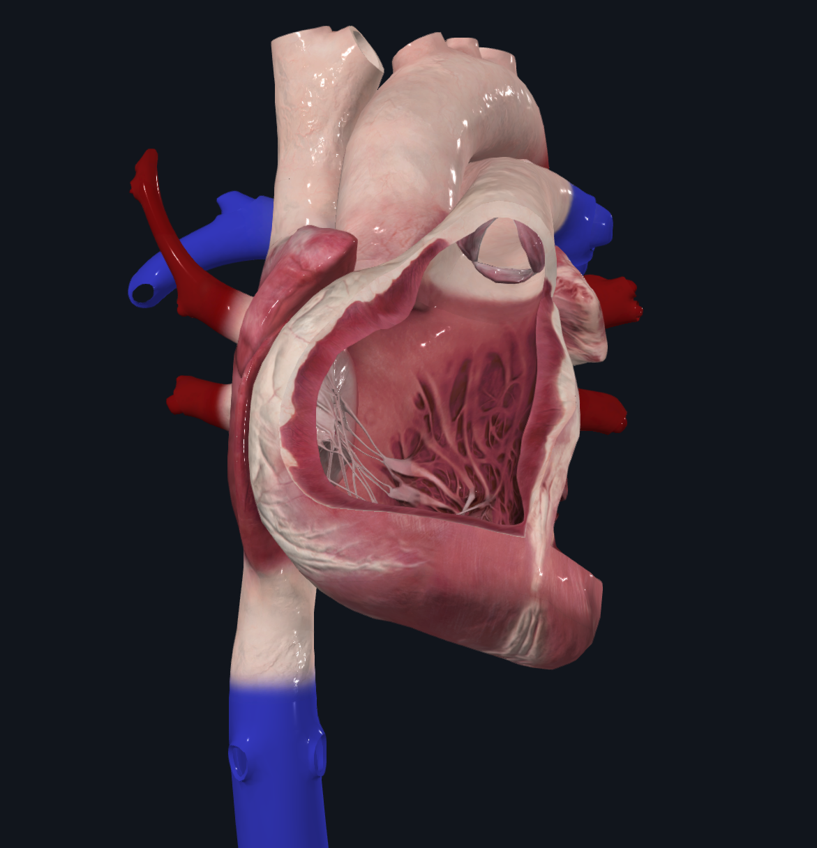
Anterior View

Explanations
- Right Atrioventricular Valve (also known as tricuspid valve)
- Allows deoxygenated blood from the right atrium to enter the right ventricles
- Chordae Tendinae
- Fibrous cords that connect the papillary muscle to the Right Atrioventricular Valve
- Papillary Muscle
- Prevents Atrioventricular valves from inverting
- Interventricular Septum
- Muscle wall that separates the right and left ventricles
- Pulmonary Semilunar Valve
- Allows deoxygenated blood to flow into the pulmonary arteries and prevents back flow
Lateral View

Explanations
- Left Atrioventricular Valve (also known as Mitral valve or bicuspid valve)
- Allows oxygenated blood from the left atrium to enter the left ventricles
- Chordae Tendinae
- Fibrous cords that connect the papillary muscle to the Left Atrioventricular Valve
- Papillary Muscle
- Prevents Atrioventricular valves from inverting
- Interventricular Septum
- Muscle wall that separates the right and left ventricles
- Aortic Semilunar Valve
- Allows oxygenatedblood to flow into the aorta and prevents back flow
Posterior view
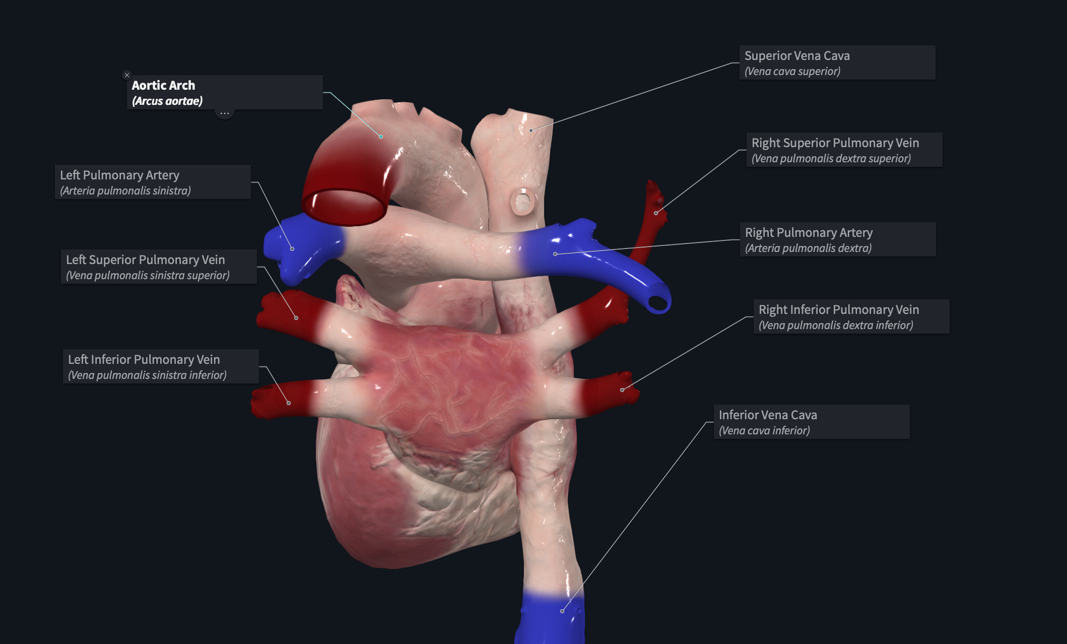
Explanations
- Aortic Arch
- Connects the ascending aorta to the descending aorta
- Superior Vena Cava
- Vertical upper chamber (anywhere above the diaphragm) for blood to enter the heart
- Inferior Vena Cava
- Vertical lower chamber (from below the diaphragm) for blood to enter the heart
- Right Pulmonary Artery
- Delivers deoxygenated blood to the right lung
- Left Pulmonary Artery
- Delivers deoxygenated blood to the left lung
- Pulmonary Veins
- Deliver oxygenated blood to the heart
- Superior means top, inferior means lower, left right mean... left and right
- Deliver oxygenated blood to the heart